A hairline crack doesn’t sound like much. But it’s often all bacteria need to get past your enamel and reach the dentin and pulp underneath. Once that happens you’re dealing with more sensitivity, more decay, and a real chance of infection or the tooth just giving out. Ignore it long enough and you’re looking at a root canal, or worse, an extraction. Catching it early is what keeps things simple: your dentist can monitor it, seal it, or restore it before it turns into a bigger, pricier problem.
Understanding Tooth Structure
A tooth is built in layers, and which layer a crack sits in changes everything about how it plays out. Enamel is the outer shell. Dentin sits underneath and carries force and sensation toward the nerve. The pulp is where the actual nerve and blood supply live. And the structures around the root hold the whole thing in place. Enamel happens to be the hardest tissue in the human body, but on the biting surface it’s only 1 to 2.5 millimeters thick. That’s not much of a buffer — damage that looks superficial can expose what’s underneath faster than people expect.
Composition of Teeth
Four parts do most of the work. Enamel is about 96% mineral by weight. Dentin is roughly 70% mineral and full of tiny tubules that carry sensation. Pulp holds the nerves, blood vessels, and immune cells. Cementum is a thin layer over the root that anchors the ligament fibers running between tooth and bone. That ligament itself is thin too, somewhere around 0.15 to 0.38 millimeters, and its job is basically shock absorption. All of this determines how much force and pain actually makes it to the nerve when something cracks.
| Layer | Details |
|---|---|
| Enamel | ~96% mineral, 1-2.5mm thick on the cusps, resists wear but can’t heal itself. |
| Dentin | ~70% mineral, has tubules that make sensitivity worse and let cracks spread. |
| Pulp | Soft tissue with nerves and blood vessels; an infection here usually means a root canal. |
| Cementum | Thin covering on the root that anchors the ligament fibers. |
| Periodontal Ligament | Connective tissue, about 0.2-0.3mm wide, that absorbs biting force. |
Types of Tooth Cracks
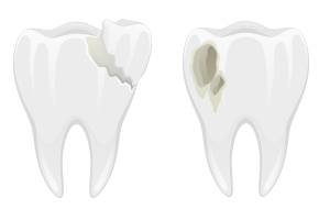
There are five you’ll typically hear about. Craze lines are superficial, mostly a cosmetic thing. A fractured cusp usually shows up near an existing filling. A cracked tooth is one where the crack has worked its way down toward the pulp. A split tooth means it’s actually separated into pieces at that point, and there’s often not much to save. A vertical root fracture starts below the gumline, which makes it the hardest to catch early. Chewing on hard things, grinding at night, or having a big filling all raise your odds. Molars take the worst of it since they absorb the most force.
- Pain that comes and goes when chewing, often right as you let go of the bite.
- Sharp, inconsistent sensitivity to cold or sweet.
- A visible line in the enamel, or a spot that feels rough that didn’t used to.
- Swelling or soreness around one particular tooth.
- Any of this showing up on and off is reason enough to get it looked at.
| Type | What It Looks Like |
|---|---|
| Craze Lines | Enamel-only, mostly cosmetic; usually no treatment needed. |
| Fractured Cusp | Often near a large filling; may need a crown or onlay to restore strength. |
| Cracked Tooth | A vertical crack that reaches into dentin, right at the pulp border; root canal plus crown are common. |
| Split Tooth | The tooth has fully separated; extraction or removing a segment is often required. |
| Vertical Root Fracture | Starts below the gumline; can cause localized bone loss and usually ends in extraction. |
Diagnosis isn’t always straightforward. Bite tests and transillumination — shining a light through the tooth — pick up somewhere between 60 and 80% of symptomatic cracks. Regular X-rays miss most vertical fractures, so a CBCT scan sometimes fills in the gap. One clinical study found that getting a crown placed promptly after a cracked-tooth diagnosis pushed three-year survival past 80%. Wait too long and the odds tilt the other way, toward pulp infection and eventually losing the tooth.
- A soft-bristled brush and steering clear of hard foods slow down any spreading.
- A night guard cuts peak biting force for people who grind, which helps too.
- Getting a large filling redone promptly spreads force more evenly.
- Talk through your options with your dentist once they’ve seen the imaging.
- Bite pain that comes and goes isn’t something to just wait out.
| Details | |
|---|---|
| Diagnosis Tools | Bite tests, transillumination, periapical X-ray, CBCT for unclear cases. |
| Conservative Treatment | A bonded onlay or crown to splint the tooth and stop the crack from spreading. |
| When a Root Canal’s Needed | Cracks reaching the pulp usually need one before any restoration. |
| When Extraction’s Needed | A split tooth or a vertical root fracture with bone loss usually can’t be saved. |
| What Affects the Outlook | How deep the crack is, where it is (crown vs. root), how fast you got treatment, and how much biting force the tooth handles. |
Causes of Small Tooth Cracks
Dietary Factors
A lot of microcracks start with what you eat and drink. Colas and sports drinks sit around pH 2.5 to 3, which is acidic enough to actively dissolve mineral out of enamel. Citrus and wine drag your mouth’s pH below the point where enamel starts to soften. Sip on something acidic for hours instead of drinking it in one go, and your saliva never gets a chance to bring things back to normal. And biting into something hard — an unpopped popcorn kernel is the classic example — puts a lot of force on one small, weak point.
- Sodas and energy drinks often sit under pH 3, which speeds up mineral loss.
- Studies show measurable softening after just a few days of frequent acid exposure.
- Acid exposure plus chewing on hard things together raise the odds of a crack a lot more than either one alone.
Dental Habits
Daily habits do a surprising amount of damage over time. Brushing hard, side to side, with a stiff-bristle brush wears down the edges of your enamel. Using your teeth to open a bottle or tear a tag puts force on them in directions they’re not built for. And grinding at night — which shows up in something like 8 to 31% of adults — applies the same repetitive stress night after night until a small crack becomes a bigger one.
Switching to a soft brush at a gentle angle helps. So does fluoride toothpaste, which strengthens spots that have already weakened. Stop using your teeth as tools. And if you grind, ask your dentist about a night guard — it takes a lot of the mechanical stress out of the equation.
Symptoms of Tooth Cracking
Sometimes it’s a hairline line you can see. Sometimes it’s a small chip, or a spot of dark discoloration. What usually gets people’s attention is sensitivity — cold, sweet, or pressure that comes and goes rather than staying constant. Pain often only shows up while chewing or right as you release your bite. A small bump or swelling in the gum nearby can mean things are heading toward infection. A molar with a vertical split, for example, can hurt sharply the moment you let go of your bite and barely at all otherwise.
Physical Indicators
Thin vertical lines, flaking enamel, or a piece of the tooth that moves slightly under pressure are all signs worth noting. Sometimes there’s staining right along the crack, or your bite just feels different than it used to. The gum next to it might get red, form a small abscess, or feel tender when you brush that specific spot.
Associated Pain
Pain here comes in two flavors. Sharp and quick, usually set off by biting something hard or letting go of pressure. Or dull and lingering, which tends to mean the pulp itself is inflamed. Cold or sweet can trigger sensitivity that lasts anywhere from a few seconds to a couple minutes, and sometimes the pain radiates into the jaw, ear, or temple — which throws people off, since it feels like the problem is somewhere else entirely.
What’s actually going on: the cracked pieces shift slightly and put stress right on the pulp, which is what causes that sharp bite pain. Bacteria can also get into the crack itself and cause pulpitis, which turns into a steady throb over the following days. There’s a case on record of someone dealing with on-and-off bite pain for weeks before the pulp finally got involved and a root canal became unavoidable — a good reminder that symptoms which come and go aren’t necessarily minor.
Risks of Ignoring Small Cracks
Leave a small crack alone and ordinary things — chewing, temperature swings — will widen it over time. What starts small can become a full fracture, an infection, or a lost tooth. Grinding, which affects up to 30% of people, speeds that timeline up considerably. Wait long enough and you’ll likely be looking at a root canal, a crown, or extraction instead of the simple bonded fix that would’ve worked earlier — plus more chair time and more money.
Progression to Larger Issues
Think of a microcrack like a fault line. Every bite, every grinding episode, makes it a little longer and a little deeper. One bad moment — biting into a pit, chewing ice — can turn a hairline crack into a full split overnight. Usually there’s a stretch of intermittent sharp pain first, a warning sign before the crack actually reaches the pulp. Once it does, infection risk climbs, and a vertical split often means extraction rather than repair.
Impact on Overall Oral Health
Once a crack opens a path to the dentin or pulp, bacteria have direct access. That raises the risk of pulpitis, an abscess, and bone loss around the root. You might end up with ongoing sensitivity, swelling, or even a small draining tract in the gum. Neighboring teeth can suffer too, since your bite shifts to avoid the problem tooth.
An infected cracked tooth usually means a root canal — one or two visits — followed by a crown. If it’s progressed to a vertical root fracture, though, extraction tends to be the only option left. Left untreated long enough, an infection can slow healing in the surrounding gum tissue too, and X-rays can pick up bone loss within months. What started as one cracked tooth can turn into a much bigger treatment plan.
Preventive Measures
Proper Oral Hygiene
Brush twice a day for two minutes with fluoride toothpaste, using a soft-bristled brush, and replace it every three months or so. Floss daily, and use interdental brushes if you’ve got wider gaps. A fluoride rinse can help if your dentist suggests one. Skip ice and hard candy, and give it about 30 minutes after anything acidic before you brush — brushing right away can actually do more damage to enamel that’s already softened.
Regular Dental Check-ups
Get checked every six months, more often if you grind or have a lot of large fillings. Bitewing X-rays every 12 to 24 months catch cracks that aren’t visible otherwise. Dentists have a few tools for this — transillumination, intraoral cameras, dye tests — that can spot a hairline fracture well before it reaches the pulp, at which point bonding, an onlay, or a crown can usually stop it in its tracks.
During a checkup, expect your dentist to check existing restorations, run bite tests, tap on suspicious teeth, and test cold sensitivity. X-rays and magnification back up what they find. If you mention pain that comes and goes with biting, or cold sensitivity, they might suggest an occlusal guard, adjust your bite, or place a protective onlay. If you grind a lot or have extensive dental work, you might get scheduled every 3 to 4 months instead of six, just so nothing gets missed.
Treatment Options
What you need depends on how deep the crack goes, what symptoms you’re dealing with, and whether the pulp’s involved. A crack that’s only in the enamel might get sealed same-day. One that’s reached the pulp usually needs a root canal and a crown to actually fix the problem and keep infection out.
Conservative Approaches
If it’s still confined to enamel or the outer dentin, you’re likely looking at bonding, a sealant, topical fluoride, an occlusal adjustment, or a night guard. Bonding and sealants are usually one visit. An occlusal adjustment might settle symptoms within a visit or two. After that, X-rays every 6 to 12 months keep tabs on whether anything’s changing.
- Dental bonding seals and stabilizes a crack in the enamel.
- Fluoride varnish or sealants help remineralize and cut sensitivity.
- An occlusal adjustment or night guard reduces the force behind grinding.
- Bitewing or periapical X-rays every 6-12 months track things over time.
| Treatment | What It Does |
|---|---|
| Dental bonding | Seals a hairline crack, restores enamel integrity, usually one visit. |
| Sealants / Fluoride | Helps remineralize and reduces sensitivity for surface-level damage. |
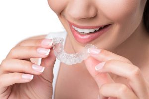
| Night guard / Occlusal adjustment | Lowers peak biting force and helps prevent the crack from spreading if you grind. |
Advanced Procedures
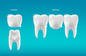
Once a crack reaches dentin or pulp, the options get more serious — a full-coverage crown, a root canal (success rates typically above 85%), or extraction followed by an implant or bridge. Crowns generally hold up for 5 to 15 years depending on material, and implants are a solid long-term answer once a tooth just can’t be saved.
Once a fracture drops below the gumline or into the root itself, things get harder to predict, and a vertical root fracture is often beyond saving — extraction becomes the likely path. Occasionally crown lengthening or periodontal surgery can rescue a borderline case, and a CBCT scan helps figure out whether surgery, restoration, or an implant makes more sense (implants run above 90% long-term success, for what it’s worth).
- A full-coverage crown protects the tooth and spreads out biting force.
- Root canal therapy removes infected pulp and keeps the tooth in place.
- Endodontic surgery (an apicoectomy) addresses issues localized to the root.
- Crown lengthening or periodontal surgery exposes more of the tooth for restoration.
- Extraction plus an implant or bridge when nothing else will save it.
| Procedure | When It’s Used |
|---|---|
| Full-coverage crown | Protects a weakened tooth; lasts roughly 5-15 years depending on material. |
| Root canal therapy | Removes infected pulp; success rates typically exceed 85%. |
| Extraction & implant | Used when the tooth can’t be saved; implants show long-term success above 90%. |
To Wrap Up
Even a tiny crack is enough of an opening for bacteria and everyday chewing forces to get past your enamel — and from there, decay, infection, sensitivity, and structural damage just keep building. Left alone, a microfracture tends to grow under normal use, and by the time it’s noticeable it’s often more complicated to fix. Getting it looked at early, while it’s still a simple problem, is what keeps you out of the root canal chair.